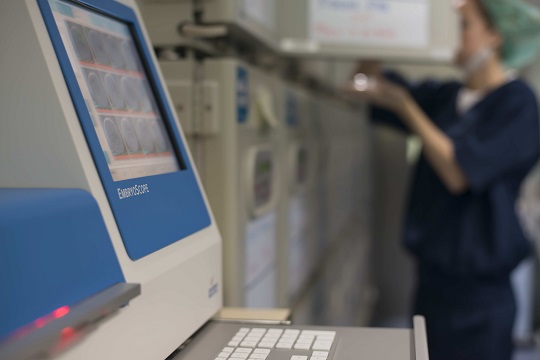

EmbryoScope allows us to record images and videos of your baby from its biological beginnings. This cutting-edge technology monitors embryo cell division in real time, capturing important information about the continuous development of the embryo. With this information, embryologists can select the healthiest embryo and the optimal time for transfer.

EmbryoScope is an incubator with an inbuilt recording device and microscope. It is capable of capturing images of embryonic development, providing a record in pictures and videos of the biological beginnings of a baby’s life. This cutting-edge technology also allows embryo cell division to be monitored in real time, which can present valuable information about the developmental health of the embryos and their potential ability to implant.
EmbryoScope™ can be used by any patient who is undergoing an assisted reproduction treatment, but it might not be suitable or beneficial for everyone. The HFEA classifies EmbryoScope as a treatment add-on, as there is not yet enough research to support its routine use for all patients but acknowledges the current research shows promise. Your consultant will assess your specific case and advise whether it is appropriate for you, allowing you to make an informed decision about whether to include EmbryoScope in your treatment plan.
EmbryoScope™ is only recommended in specific cases, including:
RESULTS
Over the last 30 years, IVI has helped more than 250,000 dreams come true.
CARE
97% of our patients recommend IVI. We’re with you at every stage of your treatment, providing support and care.
TECHNOLOGY
IVI has a worldwide reputation for innovative research and has developed and patented pioneering techniques and technologies.
EXPERTISE
IVI is one of the largest fertility treatment providers in the world, with more than 75 clinics in 9 countries.
With EmbryoScope technology, we can study of the kinetics of embryonic development. There is a relationship between the speed of cell division and embryo viability and by studying these events, it can help us select the embryos with the best potential for implantation. The time that elapses between fertilisation and the first cell division is an objective parameter and easy to determine, with predictive value in terms of embryo competence.
Traditionally, approximations for identifying the best embryos prior to transfer were based on manual monitoring conducted by an embryologist once a day. Time-lapse incubation and imaging has revolutionised the process of morphological evaluation, allowing embryology teams to study embryonic development in real-time and gain important information about their potential to implant. With this information, we can help choose the most viable embryos for transfer, improving both implantation and ongoing pregnancy rates.


By using time-lapse imaging technology, it helps embryologists determine both the healthiest embryos which are most likely to successfully implant and best time to conduct the embryo transfer procedure. For most patients, this means there is a 15 – 20% increased likelihood of a successful embryo implantation when using EmbryoScope.
The Embryoscope time-lapse system allows embryologists to monitor your embryos while the culture is performed in a stable and undisturbed environment.
The Embryoscope incubator can hold up to 6 culture plates with embryos from 6 different patients. Each culture plate can hold up to 12 embryos, so overall, the Embryoscope can take images of up to 72 embryos simultaneously.